Zeiss OCT
The Oct Allows Your Doctor to See Structures Not Visible with Only the Human Eye
What Is Optical Coherence Tomography (OCT)?
Optical coherence tomography (OCT) is a non-invasive imaging test. OCT uses light waves to take cross-section pictures of your retina.
With OCT, we can see each of the retina’s distinctive layers. This allows us to map and measure their thickness. These measurements help with diagnosis. They also provide treatment guidance for glaucoma and diseases of the retina. These retinal diseases include age-related macular degeneration (AMD) and diabetic eye disease.
What Happens During OCT?
You will sit in front of the OCT machine and rest your head on a support to keep it motionless. The equipment will then scan your eye without touching it, which only takes a few minutes.
OCT is often used to evaluate disorders of the optic nerve as well. The OCT exam helps your doctor see changes to the fibers of the optic nerve. For example, it can detect changes caused by glaucoma.
What Conditions Can OCT Help to Diagnose?
OCT is useful in diagnosing many eye conditions, including:
OCT is often used to evaluate disorders of the optic nerve as well. The OCT exam helps your eye doctor see changes to the fibers of the optic nerve. For example, it can detect changes caused by glaucoma.
OCT relies on light waves. It cannot be used with conditions that interfere with light passing through the eye. These conditions include dense cataracts or significant bleeding in the vitreous.
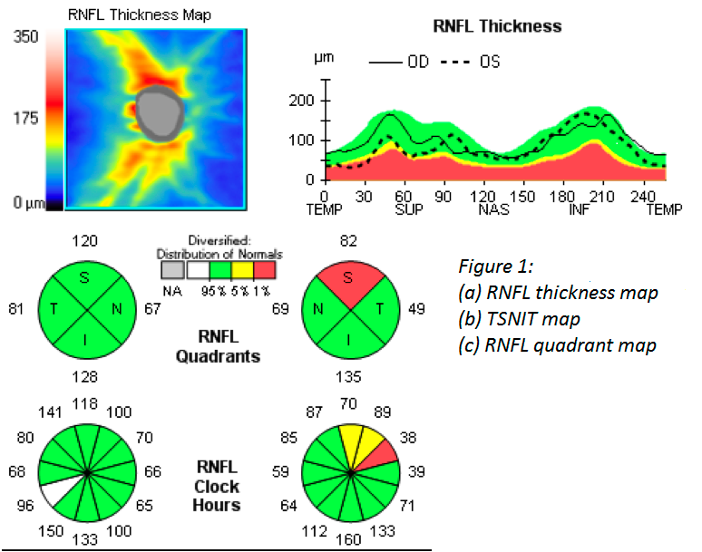
Monitoring Glaucoma with OCT
Retinal Nerve Fiber Layer (RNFL)
The RNFL is a layer of the retina. Glaucoma causes thinning of the RNFL. The TSNIT map (temporal, superior, nasal, inferior, temporal) displays RNFL thickness values by quadrants and clock hours.
Optic Nerve Head (ONH)
Think of the optic nerve like a “donut.” With uncontrolled glaucoma, the “donut hole” gets larger with time. The ONH scan quantifies the size of the “donut hole” and can pick up change before it can visually be seen.
Ganglion Cell Analysis
Similar to the RNFL, the Ganglion Cells are a layer of the retina. For patients who develop glaucoma, the Ganglion cells often become damaged before the Optic Nerve becomes damaged. In turn, when we see damage to Ganglion cells, it’s a risk factor for glaucoma.
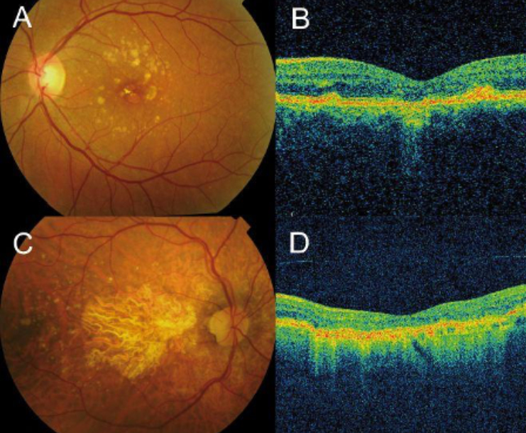
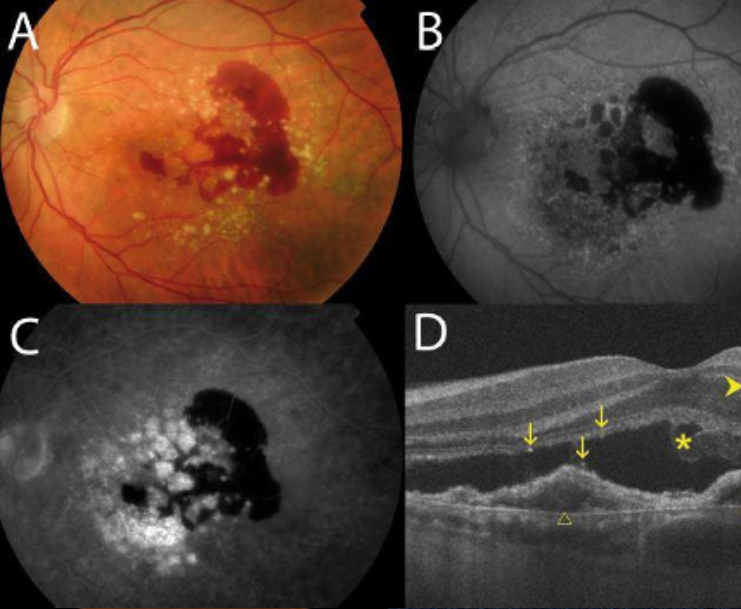
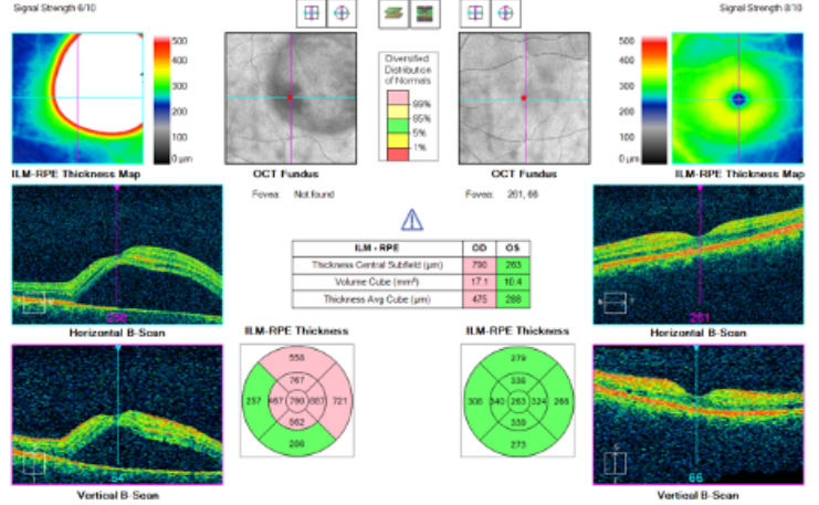
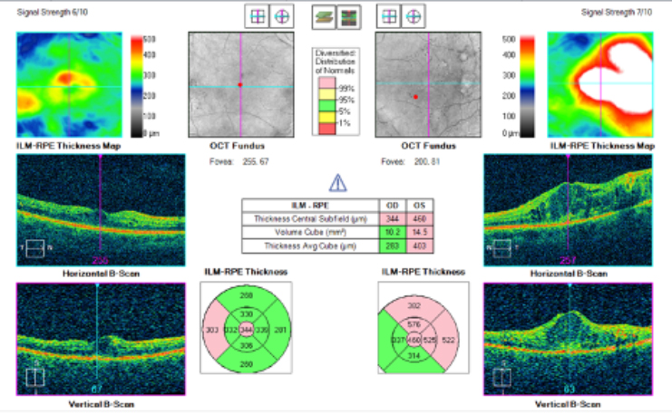
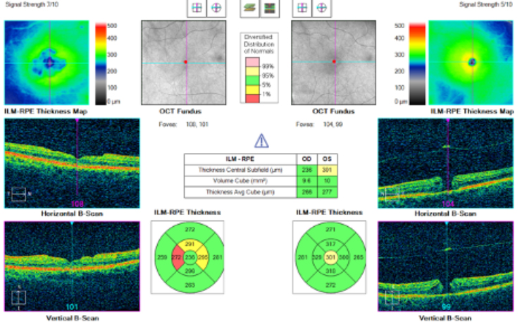
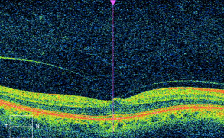
Monitoring the Macula with OCT
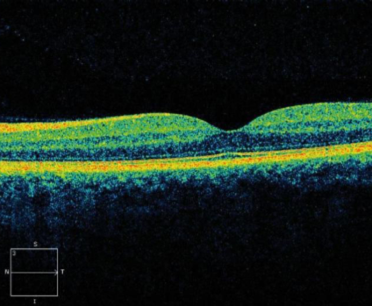
Normal Macula