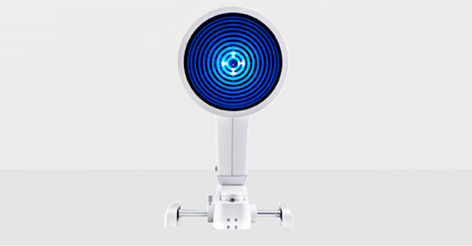
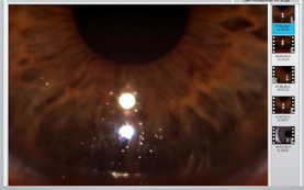
Oculus Keratograph 5M
This non-contact testing is painless and brief.
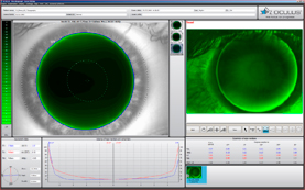
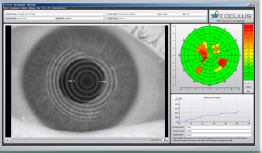
Corneal Topographer
A corneal topographer is a computer assisted diagnostic tool that creates a three-dimensional map of the surface curvature of the cornea, much like a topographic map of the earth that displays changes in the land surface. The cornea (the front window of the eye) is responsible for about 70 percent of the eye’s focusing power. An eye with normal vision has an evenly rounded cornea, but if the cornea is too flat, too steep or unevenly curved, vision will be less than perfect. The greatest advantage of corneal topography is its ability to detect irregular conditions invisible to most conventional testing.
Corneal topography produces a detailed, visual description of the shape and power of the cornea. This type of analysis provides your doctor with very fine details regarding the condition of the corneal surface. These details are used to diagnose, monitor, and treat various eye conditions. They are also used in fitting contact lenses and for planning surgery, including laser vision correction. For laser vision correction the corneal topography map is used in conjunction with other tests to determine exactly how much corneal tissue will be removed to correct vision and with what ablation pattern.
Computerized corneal topography can be beneficial in the evaluation of certain diseases and injuries of the cornea including:
- Corneal diseases
- Corneal abrasions
- Corneal deformities
- Corneal transplants
- Corneal Scars
- Irregular astigmatism following corneal transplants
- Keratoconus
- Pellucid Marginal Degeneration
- Postoperative cataract extraction with acquired astigmatism
- Pterygium
Meibo-Scan: Meibography of the Top and Bottom Eyelid
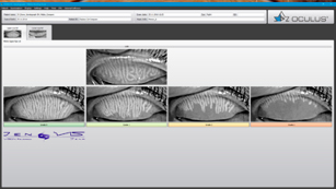
The dysfunction of meibomian glands is the most frequent cause of dry eye. Morphological changes in the gland tissue are made visible using the Meibo-Scan.
Tear Film (TF) Scan Dry Eye Workup
The TF-Scan non-invasively assesses the lipid layer and the tear film dynamics.
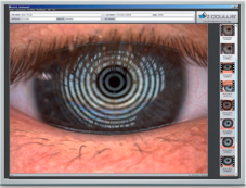
Non-Invasive Tear Break-Up Time (NITBUT)
Assessment of the Tear Film Break-Up Time
The tear film break-up time is measured non-invasively. The new infrared illumination is not visible to the human eye. This prevents glare during the examination.
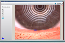
Meniscus Tear Height
Assessment of the Tear Film Quantity
The height of the tear meniscus can be precisely measured.
Lipid Layer
Assessment of the Interference Phenomenon
The interference colors of the lipid layer and their structure are made visible and can be recorded. The thickness of the lipid layer is assessed based on the structure and color.
TF Dynamics
Assessment of the Particle Flow
The video recording enables the observation of the tear film particle flow, from which conclusions regarding the viscosity of the tear film can be drawn.
Pupillometry
Pupillometry is used for the following:
- Fitting multi-focal contact lenses
- Exact determination of the treatment area for refractive surgery
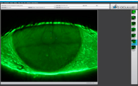
Fluorescein Imaging
Blue light makes fluorescein in tear film glow to help analyze the cornea and/or contact lens fit.